 4.9 Out of 5 based on 750+ reviews


Best Diagnostic and MRI Center in Najafgarh
This article provides an in-depth resource for patients and clinicians on diagnostic imaging services available in Najafgarh, covering MRI technology, patient preparation, service offerings, result interpretation, and practical guidance on choosing the right imaging center.
Leading Diagnostic Capabilities for Complete Medical Imaging
Najafgarh has matured into a regional healthcare hub, supported by robust diagnostic imaging infrastructure. A dependable diagnostic center is fundamental to accurate disease detection, effective treatment planning, and sustained patient care. Our diagnostic facilities combine advanced equipment, domain-experienced radiologists, and a patient-first operational model to deliver reliable imaging services for Najafgarh, Mohali, Panchkula, and surrounding areas.
We provide a broad portfolio of imaging and diagnostic modalities including MRI, CT Scan, PET-CT, Ultrasound, X-ray, Echocardiography, ECG, and EEG. Each modality is supported by modern machines and processes designed to produce accurate, reproducible results and actionable clinical reports. Our operating principle is straightforward: accurate diagnostics enable accurate treatment, and we structure our systems around speed, clarity, and clinical value.
Why Patients and Clinicians Choose Our Diagnostic Services
Selecting the appropriate diagnostic MRI Najafgarh partner has direct consequences for clinical outcomes. Below are the principal advantages that characterize our care model and explain why patients and referring clinicians prefer our centers across the Tricity.
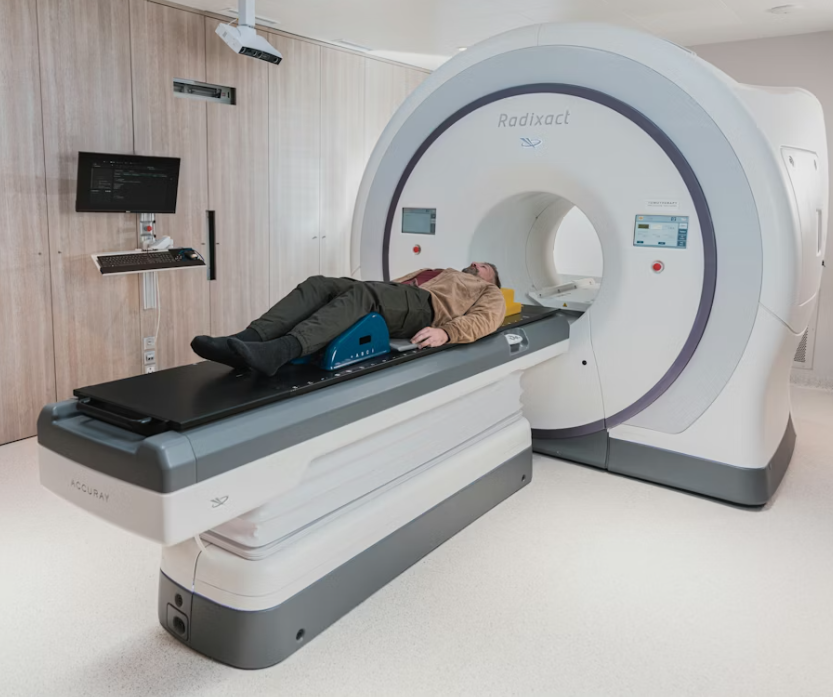
Advanced MRI Technology in Najafgarh — 1.5T & 3.0T
We operate both 1.5 Tesla and 3.0 Tesla MRI scanners to meet a broad spectrum of clinical requirements in Najafgarh. While 1.5T is suitable for routine neuro, musculoskeletal, spine, and abdominal imaging, 3.0T offers enhanced signal-to-noise and spatial resolution for complex neuroimaging, vascular assessments, oncology staging, and detailed musculoskeletal evaluations.
Accessible, Affordable Pricing
High-quality imaging should be accessible. Our pricing framework focuses on delivering premium diagnostic services at competitive rates, reducing financial barriers for families and outpatient care networks.
Same-Day Reporting by Specialist Radiologists
Reports are prepared by radiologists specializing in relevant subdisciplines — neuroradiology, musculoskeletal imaging, cardiac imaging, and abdominal imaging. Rapid turnaround supports timely clinical decisions and care planning.
Multiple, Convenient Locations
With branches in Sector 34, Sector 22, and Sector 17, our centers are positioned for convenient access across Najafgarh, Panchkula, Mohali. Workflow design focuses on short registration times and efficient patient throughput.
Patient Pick-and-Drop Assistance
To mitigate transportation challenges for elderly patients, those with mobility constraints, or individuals without private transit, we offer complimentary pick-and-drop services for select appointments.
Open Seven Days a Week
Recognizing that medical needs are not constrained to weekdays, our centers operate daily, including public holidays, ensuring appointment flexibility and urgent access when needed.
Understanding MRI: Principles and Clinical Relevance
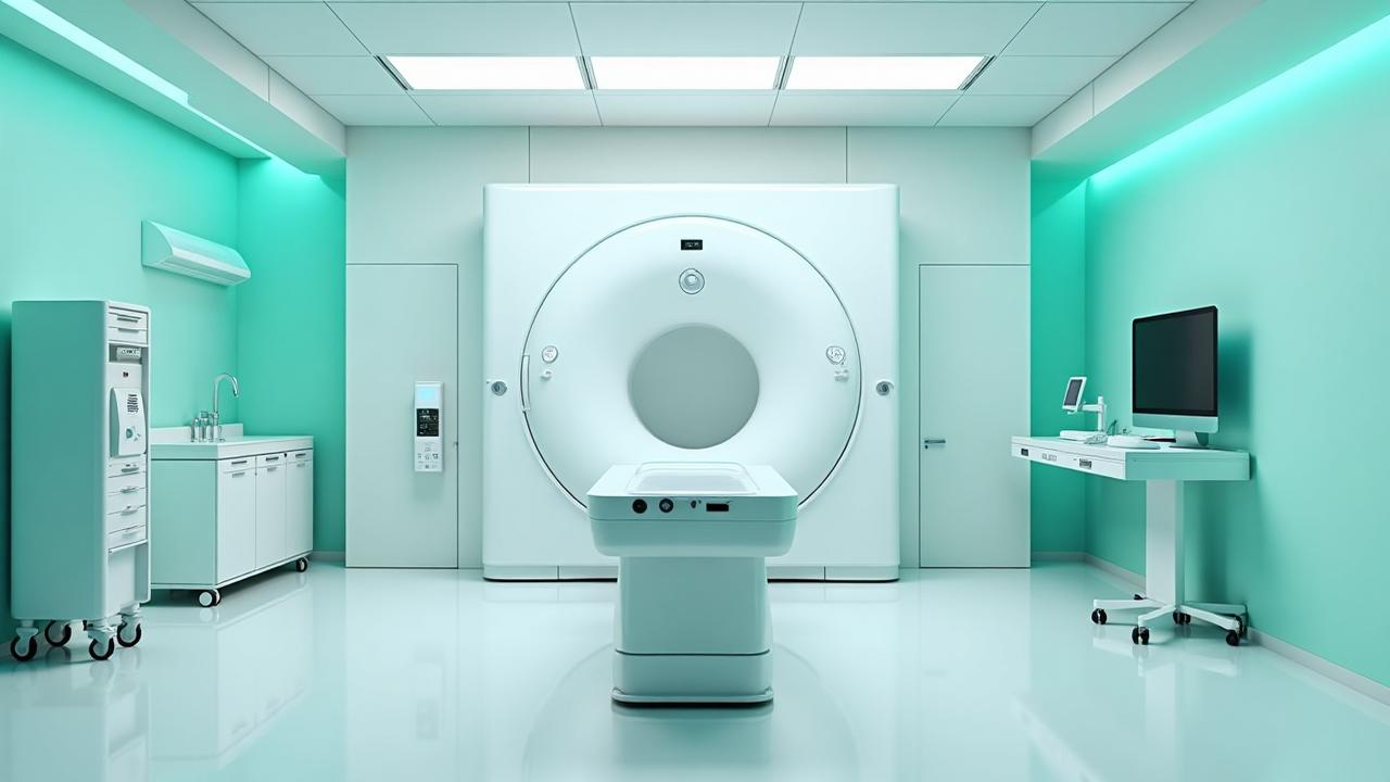
Magnetic Resonance Imaging (MRI) generates detailed images of internal structures using powerful magnetic fields and radiofrequency pulses — without ionizing radiation. MRI is particularly valuable for imaging soft tissues and complex anatomical relationships, which makes it indispensable for diagnosing a spectrum of conditions:
- Central nervous system disorders (brain and spinal cord)
- Peripheral nerve and soft-tissue pathology
- Joint and musculoskeletal injuries
- Oncologic imaging and tumor staging
- Hepatobiliary and renal assessments
- Vascular and cardiac evaluations
MRI is frequently selected where high tissue contrast is required or where radiation exposure is a concern, such as in younger patients or in repeat imaging scenarios.
1.5T versus 3.0T MRI — Clinical Implications
1.5 Tesla scanners remain the workhorse for most diagnostic needs, providing robust image quality across a wide range of applications. 3.0 Tesla scanners, by virtue of higher magnetic field strength, provide improved signal-to-noise ratio and spatial resolution, which is advantageous for:
- Detailed neuro-functional and diffusion studies
- Subtle lesion detection and early tumor identification
- Cardiac structural and functional imaging
- High-resolution musculoskeletal imaging (small joint or cartilage detail)
- Research and advanced clinical protocols
Choice of scanner is guided by clinical indication, patient safety considerations (including implants), and the diagnostic question posed by the referring physician.
Comprehensive Services Available at MRI and Diagnostic Centers in Najafgarh
Top-tier MRI centers in Najafgarh typically provide a full suite of diagnostic modalities to enable integrated assessment and efficient clinical pathways. Core services include:
Specialized MRI Protocols
Protocol-driven MRI examinations are available to address specific clinical domains:
- Brain MRI (with/without contrast; advanced sequences such as DWI, FLAIR, SWI)
- Spine MRI (cervical, thoracic, lumbar; including post-operative evaluation)
- Joint MRI (knee, shoulder, hip, wrist — including cartilage and ligament protocols)
- Whole-body MRI for systemic staging and screening in select oncology pathways
- Abdominal and pelvic MRI with liver, pancreas, and pelvic floor protocols
- Cardiac MRI and MR angiography (MRA) for vascular mapping and cardiac function
- Breast MRI for problem-solving in dense breasts or staging
Cross-Modality Capabilities
To support differential diagnosis and staging, many centers also offer:
- High-resolution CT and CT angiography
- Ultrasound and Doppler studies
- Mammography and bone densitometry
- Nuclear medicine and PET-CT for metabolic imaging
Experienced Radiology Team
Image acquisition is paired with interpretive expertise. Highly trained radiologists with subspecialty experience ensure accurate report generation, clinical correlation, and actionable recommendations. Radiology teams typically participate in multidisciplinary case reviews where imaging findings inform surgical, oncologic, and medical treatment planning.
Patient Comfort and Safety Measures
Recognizing the procedural environment of MRI (confining bore, machine noise), centers emphasize patient comfort and safety through:
- Pre-scan counselling and clear instructions
- Noise-reduction headphones and patient music options
- Cushioned supports and temperature control
- Open MRI or wide-bore options for claustrophobic or larger patients
- Continuous technician communication and panic/alert devices
- Screening for implants, allergies, renal function (for contrast), and pregnancy
Preparing for an MRI Scan — Practical Checklist
Appropriate preparation improves patient experience and image quality. The following checklist summarizes essential steps:
- Understand the procedure: Ask the center or referring clinician about expected duration, whether contrast is required, and any special positioning.
- Clothing: Wear comfortable clothing without metal fasteners. Most centers provide gowns where needed.
- Remove metal: Remove jewellery, watches, hearing aids, and any removable metallic objects before entering the scan room.
- Disclose implants: Inform staff if you have pacemakers, aneurysm clips, cochlear implants, or other metal implants; bring implant documentation if available.
- Fasting and medication: Follow specific fasting instructions for abdominal scans; continue or pause medications only per your referring physician’s advice.
- Claustrophobia or anxiety: Notify staff in advance; sedation or open MRI solutions can be arranged in many centers.
- Arrival time: Arrive early to complete paperwork and pre-scan checks.
- Documentation: Bring referral forms, prior reports/images, ID, and insurance details.
- Motion control: Remain as still as possible during scanning to avoid motion artifacts and repeat sequences.
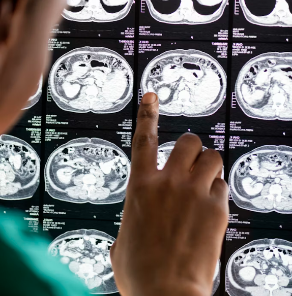
Interpreting Your MRI Results — What to Expect
After image acquisition, a radiologist examines the sequences, correlates them with clinical history, and prepares a structured report that typically contains findings, measurements, comparative statements (if prior imaging exists), and an impression summarizing clinically relevant points. Important considerations when you receive results:
- The report may include technical terminology; your referring physician will translate findings into clinical recommendations.
- Follow-up may include additional imaging, laboratory tests, or referral to a specialist depending on the findings.
- Maintain copies of reports and images for longitudinal care — these are valuable for future comparisons.
- Seek a second opinion for complex or uncertain findings; multidisciplinary review is common for oncologic and surgical planning.
Choosing a High-Quality 3T MRI Center in Najafgarh
When selecting a 3T MRI facility, consider the following quality markers:
- Availability of modern 3T and 1.5T systems and the ability to match scanner to clinical need
- Presence of subspecialty radiologists and documented reporting protocols
- Transparent pricing, insurance support, and clear pre-scan instructions
- Operational efficiency — appointment availability, minimal waiting times, and rapid report turnaround
- Infection control, facility hygiene, and accessibility (parking, transport links)
- Patient-centered amenities for comfort and anxiety mitigation
These elements collectively ensure an imaging experience that is safe, comfortable, and diagnostically robust.
About MRI Najafgarh — A Representative Example
MRI Najafgarh is one example of a comprehensive imaging provider positioned to meet diverse diagnostic needs. Centers like this emphasize integrated care: modern scanner inventory, specialist reporting, patient comfort measures, and supportive administrative processes including insurance guidance and follow-up coordination.
Located in an accessible part of the city, such centers typically serve patients from across the Tricity and beyond — balancing advanced imaging capabilities with operational convenience and patient support services.
Community-Focused Care and Clinical Partnerships
Leading diagnostic centers operate not just as imaging vendors but as clinical partners. They collaborate with hospitals, specialty clinics, and primary care providers to support screening programs, pre-operative planning, oncologic staging, and chronic disease management. Strong inter-provider communication and timely reporting are essential for integrated care pathways.
When to Ask for Advanced MRI Protocols
Request advanced imaging protocols when:
- Symptoms persist despite normal initial tests
- Subtle neurological or musculoskeletal signs require high-resolution assessment
- Oncologic staging or treatment response assessment is needed
- Detailed vascular mapping is required for surgical planning
Discuss clinical priorities with your referring clinician so the imaging center can tailor protocols to the diagnostic question.
Practical Tips for Clinicians Referring Patients
To optimize diagnostic yield and patient experience, clinicians should:
- Provide clear clinical history and focused questions on the referral
- Attach prior imaging and reports when available
- Indicate any implant or surgical history that may affect scanning
- Specify urgency to facilitate prioritized scheduling and rapid reporting when needed
Data Management, Records, and Follow-Up
Modern diagnostic centers maintain digital imaging archives and secure PACS systems that enable rapid sharing of DICOM images and electronic reports with referring physicians. Patients benefit from easy access to prior studies for comparative interpretation, and clinicians benefit from consistent, retrievable imaging records for longitudinal care.
Patient Stories and Use Cases
From acute sports injuries requiring joint imaging to complex neuro-oncology evaluations and routine cardiac assessments, MRI serves many practical use cases. Patient-centered workflows — including empathetic staff interactions, clear pre-scan counselling, and prompt report delivery — materially improve satisfaction and clinical outcomes.
Summary and Final Recommendations
High-quality diagnostic imaging in Najafgarh is characterized by modern equipment (1.5T & 3.0T MRI), subspecialty radiology expertise, patient-centered processes, transparent pricing, and integrated clinical workflows. Whether you are a patient seeking clarity about symptoms or a clinician requiring dependable imaging, select a facility that aligns scanners and protocols to specific clinical questions and provides timely, well-communicated reports.
Invest time in choosing a center that offers both advanced technology and a compassionate patient experience — the combination yields the best clinical value and supports efficient care pathways.
Next steps: Book your appointment at a center with the capabilities you need, bring prior records, and follow the patient checklist to ensure a smooth and diagnostically useful MRI examination.
For further assistance — including converting this content into a landing page, SEO-optimized blog post, or printable patient handout — tell me which format you prefer and I will prepare it.
FAQ's
1. How much does an MRI cost in Najafgarh?
The cost of an MRI in Najafgarh typically ranges between ₹2,500 to ₹7,500 depending on the type of scan, the body part, and the center. Specialized scans like 3T MRIs or contrast MRIs can cost more, up to ₹10,000.
2. Which is better, CT scan or MRI?
MRI is better for soft tissues, brain, spinal cord, joints, and tumors. CT is faster and better for bones, chest, lungs, and emergency situations. Both are complementary depending on the condition.
3. How much is a full MRI scan?
A full MRI scan of the body (whole-body MRI) can cost anywhere from ₹10,000 to ₹25,000 in Najafgarh, depending on the technology (1.5T vs 3T) and whether contrast is used.
4. What is the cost of MRI in PGI Najafgarh?
At PGIMER Najafgarh, MRI scans are subsidized for patients. The cost can be around ₹2,500–₹3,500 for general scans, but government patients often receive free or highly discounted rates.
5. Is treatment in PGI Najafgarh free?
Yes, PGIMER provides free or highly subsidized treatment to patients, especially those with government health cards or referrals, covering major surgeries, diagnostics, and therapies.
6. Can you eat before an MRI?
For most MRI scans, you can eat normally. However, for abdominal or pelvic MRIs, you may be asked to fast for 4–6 hours. Always follow the center’s instructions.
7. How long does MRI scan take?
MRI scans take 15–60 minutes depending on the body part and whether contrast is used. Whole-body MRIs may take up to 90 minutes.
8. What are two major disadvantages of MRI scans?
- High cost compared to other imaging tests.
- Claustrophobia or discomfort due to lying still in the enclosed scanner.
9. What organs are best viewed by MRI?
Brain, spinal cord, joints, heart, liver, kidneys, and soft tissue tumors are best viewed by MRI due to high soft tissue contrast.
10. Is an MRI painful or uncomfortable?
MRI is painless. Some discomfort may arise from lying still, noise, or claustrophobia. Open MRIs or sedation can help if needed.
11. Is full body MRI necessary?
Full-body MRIs are not usually necessary unless for specific screenings, high-risk patients, or research purposes. Most diagnoses require targeted scans.
12. What should I wear for an MRI?
Wear comfortable, metal-free clothing. Avoid jewelry, watches, belts, or any metal accessories. Hospitals may provide a gown for the scan.